
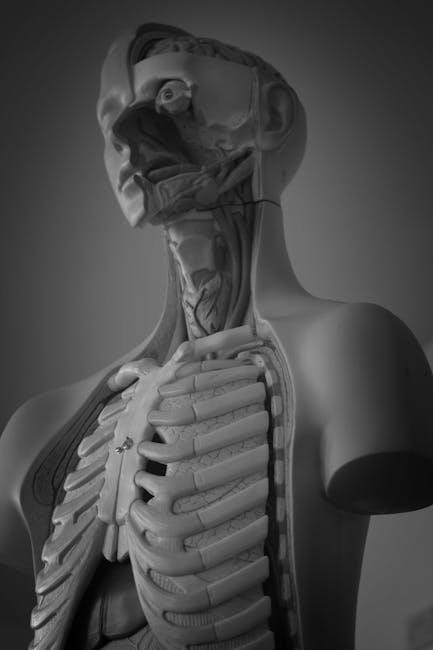
Overview of Heart Anatomy and Physiology
The heart, a vital organ, pumps blood throughout the body via the cardiovascular system. Understanding its anatomy and physiology is crucial for comprehending its function. This involves studying the heart’s structure, its chambers, valves, and blood vessels, alongside its electrical conduction and cardiac cycle.
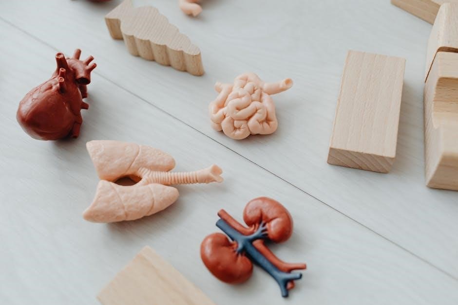
The cardiovascular system, a complex network, comprises the heart, blood vessels, and blood. This system’s primary function is to transport oxygen, nutrients, hormones, and immune cells throughout the body, while simultaneously removing waste products like carbon dioxide. The heart, acting as the central pump, propels blood through arteries, veins, and capillaries, ensuring efficient circulation.
The lymphatic system, closely associated, aids in fluid balance and immunity. A thorough understanding of the heart’s anatomy and physiology is essential for comprehending the entire cardiovascular system’s intricate workings. From the rhythmic contractions orchestrated by the sinoatrial node to the precise opening and closing of heart valves, each component plays a vital role in maintaining life.
Disruptions in the system can cause heart diseases, therefore, it is very important to study the heart’s anatomy and physiology. This system helps maintain balance within the body.

Heart Anatomy
Heart anatomy encompasses the physical structures of the heart. This includes the heart’s location, layers, chambers, and valves. Studying these anatomical features is crucial to understanding how the heart functions as a pump within the cardiovascular system;
Location and Orientation of the Heart
The heart resides within the thorax, centrally positioned, and slightly tilted. Its apex, formed by the left ventricle, points downward, forward, and to the left, typically at the 5th left intercostal space. This unique orientation is vital for efficient blood pumping.
The heart is surrounded by the pericardium and situated between the lungs in the mediastinum. Its location is crucial for its protection and interaction with surrounding structures. Superiorly, major blood vessels connect, while inferiorly, it rests on the diaphragm.
Understanding its precise location and orientation is fundamental for medical professionals. This knowledge is essential for accurate diagnosis and treatment of cardiac conditions, including physical examinations and imaging techniques. The heart’s placement is also crucial during surgical procedures. Knowledge of the heart’s position helps to ensure accurate and safe interventions.
Layers of the Heart Wall
The heart wall comprises three distinct layers: the epicardium, myocardium, and endocardium, each with specific functions. The epicardium, the outermost layer, is also the visceral layer of the serous pericardium. This layer contains blood vessels and nerves, providing nourishment and control to the heart.
The myocardium, the thickest layer, consists of cardiac muscle responsible for the heart’s pumping action. Its contractions propel blood throughout the circulatory system. The arrangement of myocardial fibers is complex, optimizing the heart’s ability to generate force efficiently.
The endocardium, the innermost layer, lines the heart chambers and covers the valves. It is continuous with the endothelium of blood vessels, ensuring a smooth surface for blood flow. This layer is essential for preventing blood clot formation within the heart. These layers work together to maintain cardiac function.
Chambers of the Heart
The human heart consists of four chambers: two atria and two ventricles. The atria, the upper chambers, receive blood returning to the heart. The right atrium receives deoxygenated blood from the body via the superior and inferior vena cava, while the left atrium receives oxygenated blood from the lungs through the pulmonary veins.
The ventricles, the lower chambers, are responsible for pumping blood out of the heart. The right ventricle pumps deoxygenated blood to the lungs via the pulmonary artery for oxygenation. The left ventricle, the largest and strongest chamber, pumps oxygenated blood to the entire body through the aorta.
These chambers function in a coordinated manner to ensure efficient blood circulation. The atria contract to fill the ventricles, which then contract to pump blood out of the heart, maintaining the circulatory system’s continuous flow.
Valves of the Heart
The heart’s efficient function relies heavily on its valves, which ensure unidirectional blood flow through the chambers. There are four main valves: the tricuspid, mitral (bicuspid), pulmonary, and aortic valves.
The tricuspid valve, located between the right atrium and right ventricle, prevents backflow of blood into the right atrium during ventricular contraction. The mitral valve, positioned between the left atrium and left ventricle, similarly prevents backflow into the left atrium.
The pulmonary valve, situated between the right ventricle and the pulmonary artery, ensures blood flows only towards the lungs. The aortic valve, located between the left ventricle and the aorta, prevents backflow of blood into the left ventricle.
These valves open and close in coordination with the heart’s contractions and relaxations, ensuring blood moves forward with each heartbeat. Valve dysfunction can lead to various heart conditions, affecting the circulatory system’s overall efficiency.
Coronary Arteries and Blood Supply
The heart, a tireless pump, requires a constant supply of oxygen and nutrients to function optimally. This crucial task is performed by the coronary arteries, a network of blood vessels that surround and penetrate the heart muscle.
The two main coronary arteries, the left and right coronary arteries, originate from the aorta, just above the aortic valve. The left coronary artery typically branches into the left anterior descending artery and the circumflex artery, supplying blood to the left ventricle and left atrium.
The right coronary artery supplies blood to the right ventricle, right atrium, and the posterior part of the left ventricle in most individuals. These arteries branch further into smaller vessels, ensuring that all parts of the heart receive adequate blood flow.
Blockage or narrowing of the coronary arteries, often due to atherosclerosis, can lead to reduced blood flow, causing chest pain (angina) or even a heart attack (myocardial infarction).
Heart Physiology
Heart physiology delves into the mechanics of cardiac function. It covers the cardiac cycle, electrical conduction, and cardiac output, all essential for understanding how the heart efficiently pumps blood throughout the body. These intricate processes are vital for life.
Cardiac Cycle
The cardiac cycle encompasses the coordinated sequence of events that occur during one complete heartbeat. It includes diastole, where the heart chambers relax and fill with blood, and systole, where the chambers contract and eject blood into the pulmonary and systemic circulations. This cycle relies on pressure gradients, ensuring unidirectional blood flow through the heart’s chambers and valves.
During diastole, the atria fill with blood returning from the body and lungs, while the ventricles passively fill. Atrial contraction then actively propels additional blood into the ventricles, maximizing their filling capacity. Systole begins with ventricular contraction, increasing pressure and closing the atrioventricular valves (mitral and tricuspid), preventing backflow into the atria. As ventricular pressure rises further, it exceeds the pressure in the aorta and pulmonary artery, causing the semilunar valves to open and blood to be ejected.
The cardiac cycle is tightly regulated, ensuring that the heart efficiently pumps blood to meet the body’s metabolic demands. Factors like heart rate, contractility, and preload (ventricular filling) influence the cycle’s duration and the volume of blood ejected with each beat.
Electrical Conduction System of the Heart

The heart’s electrical conduction system is a specialized network of cells responsible for initiating and coordinating heart muscle contractions. This system ensures a rhythmic and synchronized heartbeat, essential for efficient blood pumping. The sinoatrial (SA) node, located in the right atrium, serves as the heart’s natural pacemaker.
The SA node generates electrical impulses that spread throughout the atria, causing them to contract. These impulses then reach the atrioventricular (AV) node, which briefly delays the signal, allowing the atria to fully contract before ventricular activation. From the AV node, the signal travels down the bundle of His, a specialized pathway that divides into left and right bundle branches.
These bundle branches conduct the impulses to the Purkinje fibers, a network of fibers that spread throughout the ventricular myocardium. The Purkinje fibers rapidly transmit the electrical signal, causing the ventricles to contract in a coordinated manner. This intricate system ensures that the heart beats rhythmically and efficiently, delivering oxygenated blood to the body.
Cardiac Output and its Regulation
Cardiac output (CO) is the volume of blood pumped by the heart per minute, a crucial indicator of cardiovascular function. It’s calculated by multiplying heart rate (HR), the number of beats per minute, by stroke volume (SV), the amount of blood ejected with each beat. CO reflects the heart’s ability to meet the body’s oxygen and nutrient demands.
Several factors regulate CO. Heart rate is influenced by the autonomic nervous system, with sympathetic stimulation increasing HR and parasympathetic stimulation decreasing it. Stroke volume is determined by preload, afterload, and contractility. Preload, the volume of blood in the ventricles at the end of diastole, increases SV.
Afterload, the resistance the heart must overcome to eject blood, decreases SV. Contractility, the force of ventricular contraction, increases SV. The body regulates CO through neural and hormonal mechanisms to maintain adequate tissue perfusion. Understanding CO and its regulation is essential for assessing cardiovascular health and diagnosing heart conditions.
Be First to Comment